Undergraduate Research at Jefferson Lab
Development of a Scintillation Web Detector for Real-Time Radioisotope Imaging of 32P Uptake in the Root Systems of Plants
Student: Anthony Llodra
School: Florida International University
Mentored By: Drew Weisenberger, Brian Kross, Carl Zorn and Seung Joon Lee
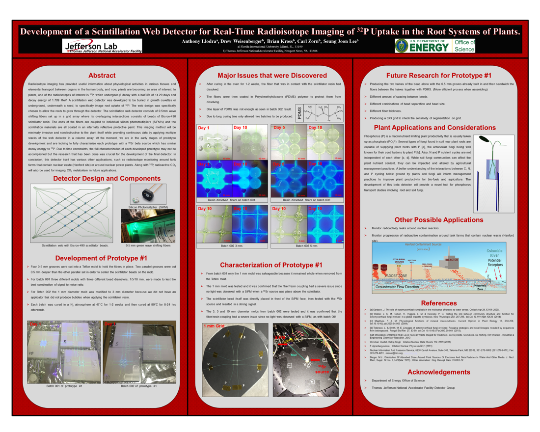
Radioisotope imaging has provided useful information about physiological activities in various tissues and elemental transport between organs in the human body, and now, plants are becoming an area of interest. In plants, one of the radioisotopes of interest is 32P. A scintillation web detector was developed to be buried in growth cuvettes or underground, underneath a seed, to specifically image root uptake of 32P. The web design was specifically chosen to allow the roots to grow through the detector. The scintillation web detector consists of a grid array of 0.5mm wave shifting fibers where it's overlapping intersections consists of beads of Bicron-490 scintillator resin. The ends of the fibers are coupled to individual silicon photomultipliers (SiPM's) and the scintillation materials are all coated in an internally reflective protective paint. This imaging method will be minimally invasive and nondestructive to the plant itself while providing continuous data by applying multiple stacks of the web detector in a column array. At the moment, we are in the early stages of prototype development and the development of prototype #1 followed by characterization of the prototype with a 90Sr beta source, which has similar decay energy to 32P. Due to time constraints, the full characterization of prototype #1 may not be accomplished but the research that has been done was crucial for the development of the final detector. In conclusion, this detector itself has various other applications, such as radioisotope monitoring around tank farms that contain nuclear waste (Hanford site) or around nuclear power plants.

Citation and linking information
For questions about this page, please contact Education Web Administrator.
